Improving Vertebrate Skeleton Images: Fluorescence and the Non-Permanent Mounting of Cleared-and-Stained Specimens
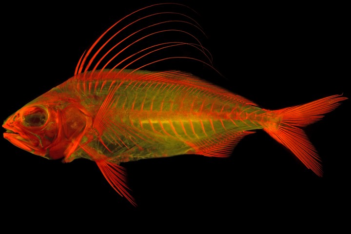 Image credit: Matthew G. Girard
Abstract
Visualizing complex morphological features using digital photographs is often challenging in comparative anatomical studies. Progress in comparative anatomical studies has made substantive shifts through the development of new and improved methods for preparing specimens and visualizing characters. The advent of enzyme-cleared and bone-stained specimens revolutionized comparative anatomical studies in the middle of the 20th century. Continued refinement and improvement on these techniques combined with alternative approaches to visualization have allowed for more detailed investigations of vertebrate anatomy. One of the most difficult challenges remaining in comparative anatomy is accurately communicating morphological variation, and methodological improvements that refine the visual explanation are critical. Here we present two methods that simplify and improve the digital imaging of vertebrate skeletons and their components. First, fluorescence microscopy with alizarin-stained specimens is shown to help identify bony margins, facilitate the identification of skeletal elements in extant and fossil specimens, enhance the light alizarin staining of bone, and differentiate skeletal and soft tissues. Second, the non-permanent mounting of cleared- and-stained vertebrate specimens in a glycerine-gelatin matrix allows researchers to temporarily pose specimens for otherwise impossible scientific or artistic images. These two methods greatly improve researchers’ ability to visualize vertebrate specimens or characters they are describing. The improved communication of critical anatomical variation through visual means facilitates the explanations demanded by evolutionary research, specifically, and biology, generally.
Almetric and Dimension badges